The following are descriptions of several imaging research projects that our medical physicists are currently pursuing:
Dual Energy Computed Tomography for Bone Delineation (Lawless)
Using the tools available to us as a part of the Siemens Somatom Definition Edge Dual Energy CT (DECT) scanner, we have the ability to better visualize tissues that are historically difficult to differentiate. With this technology, we are working toward being able to delineate the red and yellow bone marrow present near a patient’s treatment site. Additionally, we are investigating the radiation response of bone marrow to doses in the therapeutic range, so as to be able to predict any effect on a patient’s hematopoetic system. Through the creation of virtual non-calcium images we can better visualize abnormalities within bony structures, which will increase the visibility of bone lesions. The research project is founded in optimizing the algorithm parameters for our purposes and leveraging the system to perform optimally for these purposes.
Cone Beam Computed Tomography Dose Investigation (Lawless)
The dose from pre-treatment cone beam computed tomography scans has been investigated extensively, but most assessments focus on the in-field dose. In certain instances, such as the treatment of pregnant patients, the out-of-field dose from imaging procedures could contribute a significant dose to areas of concern (such as the fetus). Measuring this dose can prove difficult, and this research will investigate which radiation detectors can and should be used for these purposes and how to calibrate them. The dose information gathered from this investigation will be used to inform decisions on imaging protocols for clinical patients.
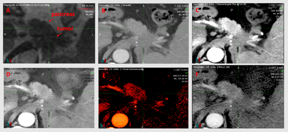
Improving Tumor Delineation in the Pancreas and Liver Through Dual-Energy CT (Miller)
Tumor delineation in the pancreas and liver can be a challenge using conventional CT images. Dual-Energy CT provides many opportunities to better delineate tumor from healthy tissue and therefore has great potential to aid in radiation therapy. The Department of Human Oncology has installed a novel single-source dual-energy CT system, called TwinBeam, with potential for liver and pancreas imaging. We are currently quantifying the advantages gained through TwinBeam dual-energy CT.